
Two Cases of Skin Mycosis Due to Hanseniaspora Opuntiae and Cutaneotrichosporon Mucoides at Basrah Southern of Iraq
DOI:
https://doi.org/10.48112/bcs.v2i3.558Abstract
 Abstract Views: 737
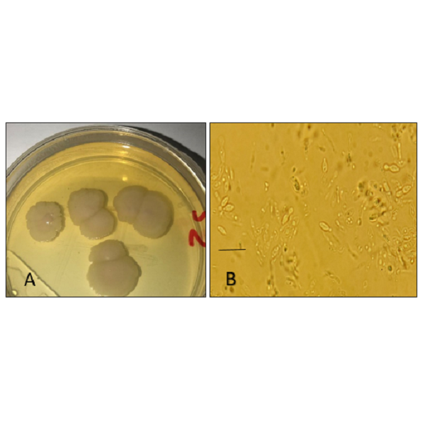
Mycoses of the skin are a group of infections that affect the skin and its appendages, mainly caused by dermatophytic fungi, and may be caused by yeasts or rearly by non-dermatophytic molds. Skin swabs collected from 60 patients attending the Dermatology Clinics at Madinah Central Hospital and Al-Fayhaa General Hospital in Basrah province from October 2021 to March 2022 were surveyed for the presence of dermatomycoses. Direct microscopical examination was carried out with 15% KOH, and repeated cultures were performed on Sabouraud dextrose agar with chloramphenicol showed the same colonies. In this paper,we present two interesting cases in which Hanseniaspora opuntiae HAM17 and Cutaneotrichosporon mucoides HAM14 can be distinctly identified as causative agent of cutaneous mycoses. In the first case, we describe a new etiologic agent, Hanseniaspora opuntiae HAM17, which was implicated in a cutaneous infection in a 45-year-old woman with a history of diabetes mellitus, and to the best of our knowledege, it represents the fourth clinical case due to this fungus in the world. The second case involved cutaneous mycosis due to Cutaneotrichosporon mucoides HAM14 in a 23-year-old woman showed inflammatory lesions similar to acne on the back, and she was suffering from hormonal disorders. This case and the etiologic agent are reported for the first time in Iraq. The isolated yeast species were examined and purified for phenotypic identification and genetical analysis using the primers ITS1-ITS4. Sequences were deposited into Japanese Genbank as new strains under accession numbers LC722487 and LC722484.
Keywords:
Case report in Basrah, Cutaneotrichosporon mucoides, Hanseniaspora opuntiae, Molecular identification, Skin mycosisReferences
Arastehfar, A., Daneshnia, F., Farahyar, S., Fang, W., Salimi, M., Salehi, M., ... & Boekhout, T. (2019). Incidence and spectrum of yeast species isolated from the oral cavity of Iranian patients suffering from hematological malignancies. Journal of Oral Microbiology, 11(1), 1601061. https://doi.org/10.1080/20002297.2019.1601061
Basu, S., Prakash, K., Gugnani, H. C., Joshi, S., Wattal, C., & Padhye, A. A. (2003). Isolation of Trichosporon mucoides from urine. Journal de Mycologie Medicale, 13(3), 155-156.
Basu, S., Tilak, R., & Kumar, A. (2015). Multidrug-resistant Trichosporon: an unusual fungal sepsis in preterm neonates. Pathogens and Global Health, 109(4), 202-206. https://doi.org/10.1179/2047773215Y.0000000019
Cadez, N., Poot, G. A., Raspor, P., & Smith, M. T. (2003). Hanseniaspora meyeri sp. nov., Hanseniaspora clermontiae sp. nov., Hanseniaspora lachancei sp. nov. and Hanseniaspora opuntiae sp. nov., novel apiculate yeast species. International Journal of Systematic and Evolutionary Microbiology, 53(5), 1671-1680. https://doi.org/10.1099/ijs.0.02618-0
Chen, H., Zhou, X., Ren, B., & Cheng, L. (2020). The regulation of hyphae growth in Candida albicans. Virulence, 11(1), 337-348. https://doi.org/10.1080/21505594.2020.1748930
Chen, Y. T., Yang, W. C., Chen, T. W., & Lin, C. C. (2013). Trichosporon mucoides peritonitis in a continuous ambulatory peritoneal dialysis patient. Peritoneal Dialysis International, 33(3), 341-342. https://doi.org/10.3747/pdi.2012.00146
Colombo, A. L., Padovan, A. C. B., & Chaves, G. M. (2011). Current knowledge of Trichosporon spp. and Trichosporonosis. Clinical microbiology reviews, 24(4), 682-700. https://doi.org/10.1128/cmr.00003-11
de Almeida Júnior, J. N., & Hennequin, C. (2016). Invasive Trichosporon infection: a systematic review on a re-emerging fungal pathogen. Frontiers in Microbiology, 7, 1629. https://doi.org/10.3389/fmicb.2016.01629
De Hoog, G. S., Guarro, J., Gene, J., & Figueras, M. J. (2000). Atlas of clinical fungi, Centraalbureau voor Schimmelcultures. Utrecht, the Netherlands, 276-282.
Ebert, A., Monod, M., Salamin, K., Burmester, A., Uhrlaß, S., Wiegand, C., ... & Nenoff, P. (2020). Alarming India‐wide phenomenon of antifungal resistance in dermatophytes: a multicentre study. Mycoses, 63(7), 717-728. https://doi.org/10.1111/myc.13091
Eddouzi, J., Lohberger, A., Vogne, C., Manai, M., & Sanglard, D. (2013). Identification and antifungal susceptibility of a large collection of yeast strains isolated in Tunisian hospitals. Medical Mycology, 51(7), 737-746. https://doi.org/10.3109/13693786.2013.800239
Ellis, D. H. (1994). Clinical mycology: The human opportunistic mycoses. Pfizer Incorporated.
Gnat, S., Łagowski, D., Dyląg, M., & Nowakiewicz, A. (2021). European hedgehogs (Erinaceus europaeus L.) as a reservoir of dermatophytes in Poland. Microbial ecology, 1-13. https://doi.org/10.1007/s00248-021-01866-w
González-Abad, M. J. (2018). Isolation of Hanseniaspora opuntiae from blood cultures of an immunosuppressed patient: An infrequent and difficult to assess finding. Revista Iberoamericana de Micologia, 35(3), 168-169. https://doi.org/10.1016/j.riam.2018.05.001
Guého, E., Improvisi, L., De Hoog, G. S., & Dupont, B. (1994). Trichosporon on humans: A practical account: Trichosporon am Menschen: Eine Darstellung für die Praxis. Mycoses, 37(1‐2), 3-10. https://doi.org/10.1111/j.1439-0507.1994.tb00277.x
Guého, E., Smith, M. T., De Hoog, G. S., Billon-Grand, G., Christen, R., & van der Vegte, W. B. (1992). Contributions to a revision of the genus Trichosporon. Antonie van Leeuwenhoek, 61, 289-316. https://doi.org/10.1007/BF00713938
Gupta, A. K., & Cooper, E. A. (2008). Update in antifungal therapy of dermatophytosis. Mycopathologia, 166(5-6), 353-367. https://doi.org/10.1007/s11046-008-9109-0
Hasan, A. M. (2014). The effect of some antifungals on fungal species that isolated from Leukemia patients. Athesis. College of Science, University of Baghdad.
Hindy, N., & Abiess, A. A. (2019). Isolation and identification of dermatophytes causing Dermatophytosis in Hilla city, Iraq. Indian Journal of Public Health Research & Development, 10(10), 2225-2230.
Kannan, P., Janaki, C., & Selvi, G. S. (2006). Prevalence of dermatophytes and other fungal agents isolated from clinical samples. Indian Journal of Medical Microbiology, 24(3), 212-215. https://doi.org/10.1016/S0255-0857(21)02353-7
Kauffman, S. A. (2000). Investigations. Oxford University Press.
Kendirli, T., Ciftçi, E., Ince, E., Öncel, S., Dalgiç, N., Güriz, H., ... & Dogru, U. (2006). Successful treatment of Trichosporon mucoides infection with lipid complex amphotericin B and 5‐fluorocytosine. Mycoses, 49(3), 251-253. https://doi.org/10.1111/j.1439-0507.2006.01223.x
Lakshmanan, A., Ganeshkumar, P., Mohan, S. R., Hemamalini, M., & Madhavan, R. (2015). Epidemiological and clinical pattern of dermatomycoses in rural India. Indian Journal of Medical Microbiology, 33, S134-S136. https://doi.org/10.4103/0255-0857.150922
Lee, W. J., Kim, J. Y., Song, C. H., Jung, H. D., Lee, S. J., & KIM, D. W. (2011). Disruption of barrier function in dermatophytosis and pityriasis versicolor. The Journal of Dermatology, 38(11), 1049-1053. https://doi.org/10.1111/j.1346-8138.2011.01320.x
Liu, X. Z., Wang, Q. M., Göker, M., Groenewald, M., Kachalkin, A. V., Lumbsch, H. T., ... & Bai, F. Y. (2015). Towards an integrated phylogenetic classification of the Tremellomycetes. Studies in Mycology, 81(1), 85-147. https://doi.org/10.1016/j.simyco.2015.12.001
Lopes, G., Pinto, E., & Salgueiro, L. (2017). Natural products: an alternative to conventional therapy for dermatophytosis?. Mycopathologia, 182, 143-167. https://doi.org/10.1007/s11046-016-0081-9
Luan, Y., Zhang, B. Q., Duan, C. Q., & Yan, G. L. (2018). Effects of different pre-fermentation cold maceration time on aroma compounds of Saccharomyces cerevisiae co-fermentation with Hanseniaspora opuntiae or Pichia kudriavzevii. LWT, 92, 177-186. https://doi.org/10.1016/j.lwt.2018.02.004
Miron, D., Battisti, F., Silva, F. K., Lana, A. D., Pippi, B., Casanova, B., ... & Schapoval, E. E. (2014). Antifungal activity and mechanism of action of monoterpenes against dermatophytes and yeasts. Revista Brasileira de farmacognosia, 24, 660-667. https://doi.org/10.1016/j.bjp.2014.10.014
Murray, P. R., Rosenthal, K. S., & Pfaller, M. A. (2020). Medical microbiology E-book. Elsevier Health Sciences, 9, 426-433.
Nishiura, Y., Nakagawa-Yoshida, K., Suga, M., Shinoda, T., Guého, E., & Ando, M. (1997). Assignment and serotyping of Trichosporon species: the causative agents of summer-type hypersensitivity pneumonitis. Journal of Medical and Veterinary Mycology, 35(1), 45-52. https://doi.org/10.1080/02681219780000861
Nisiotou, A. A., & Nychas, G. J. E. (2007). Yeast populations residing on healthy or Botrytis-infected grapes from a vineyard in Attica, Greece. Applied and Environmental Microbiology, 73(8), 2765-2768. https://doi.org/10.1128/AEM.01864-06
Oh, T. H., Shin, S. U., Kim, S. S., Kim, S. E., Kim, U. J., Kang, S. J., ... & Park, K. H. (2020). Prosthetic valve endocarditis by Trichosporon mucoides: A case report and review of literature. Medicine, 99(41). https://doi.org/10.1097%2FMD.0000000000022584
Oliveira dos Santos, C., Zijlstra, J. G., Porte, R. J., Kampinga, G. A., van Diepeningen, A. D., Sinha, B., & Bathoorn, E. (2016). Emerging pan-resistance in Trichosporon species: a case report. BMC Infectious Diseases, 16(1), 1-4. https://doi.org/10.1186/s12879-016-1477-3
Padhi, S., Dash, M., Pattanaik, S., & Sahu, S. (2014). Fungemia due to Trichosporon mucoides in a diabetes mellitus patient: a rare case report. Indian journal of medical microbiology, 32(1), 72-74. https://doi.org/10.4103/0255-0857.124324
Papalexandratou, Z., Lefeber, T., Bahrim, B., Lee, O. S., Daniel, H. M., & De Vuyst, L. (2013). Hanseniaspora opuntiae, Saccharomyces cerevisiae, Lactobacillus fermentum, and Acetobacter pasteurianus predominate during well-performed Malaysian cocoa bean box fermentations, underlining the importance of these microbial species for a successful cocoa bean fermentation process. Food Microbiology, 35(2), 73-85. https://doi.org/10.1016/j.fm.2013.02.015
Pierre, Y., Christopher, S., Namoomba, S. H., Lukwesa, M., & Kwenda, G. (2016). Isolation and Identification of fungi from suspected fungal skin infections in patients attending the Dermatology Clinic at University Teaching Hospital IOSR Journal Of Pharmacy. Issue, 10, 55-61.
Sambrook, J., Fritsch, E. F., & Maniatis, T. (2012). Molecular Cloning: A Laboratory Manual, 4 Edn New York. NY: Cold Spring Harbor Laboratory Press.
Seebacher, C., Bouchara, J. P., & Mignon, B. (2008). Updates on the epidemiology of dermatophyte infections. Mycopathologia, 166, 335-352. https://doi.org/10.1007/s11046-008-9100-9
Theelen, B., Cafarchia, C., Gaitanis, G., Bassukas, I. D., Boekhout, T., & Dawson Jr, T. L. (2018). Malassezia ecology, pathophysiology, and treatment. Medical mycology, 56(suppl_1), S10-S25. https://doi.org/10.1093/mmy/myx134
Tse, C., Boodman, C., & Wuerz, T. (2022). Trichosporon mucoides prosthetic valve endocarditis managed with antifungal suppression therapy. Medical Mycology Case Reports, 36, 10-12. https://doi.org/10.1016/j.mmcr.2022.02.004
Vallabhaneni, S., Kallen, A., Tsay, S., Chow, N., Welsh, R., Kerins, J., ... & Chiller, T. M. (2016). Investigation of the first seven reported cases of Candida auris, a globally emerging invasive, multidrug-resistant fungus—United States, May 2013–August 2016. Morbidity and Mortality Weekly Report, 65(44), 1234-1237. https://www.jstor.org/stable/24859138
Weeks, J. E. F. F., Moser, S. A., & Elewski, B. E. (2003). Superficial cutaneous fungal infections. Clinical Mycology, 367.
White, T. J., Bruns, T., Lee, S. J. W. T., & Taylor, J. (1990). Amplification and direct sequencing of fungal ribosomal RNA genes for phylogenetics. PCR protocols: a guide to methods and applications, 18(1), 315-322.
Zimbres, A. C. G., Albuquerque, P. C., Joffe, L. S., Souza, T. N., Nimrichter, L., Frazão, S. O., ... & Rodrigues, M. L. (2018). A glucuronoxylomannan-like glycan produced by Trichosporon mucoides. Fungal Genetics and Biology, 121, 46-55. https://doi.org/10.1016/j.fgb.2018.09.009

Downloads
Published
How to Cite
Issue
Section
License
Copyright (c) 2023 Biomedicine and Chemical Sciences

This work is licensed under a Creative Commons Attribution 4.0 International License.


